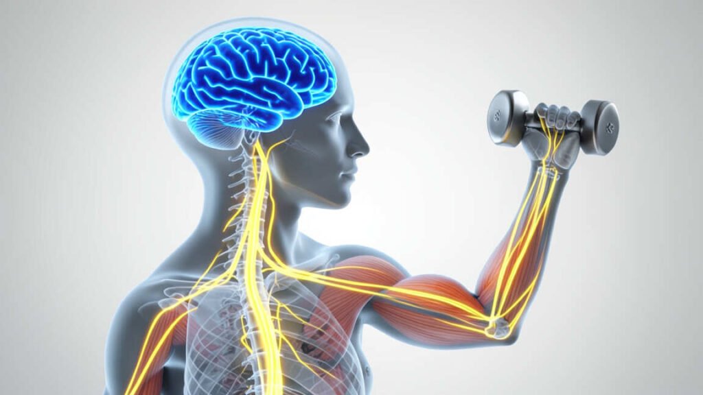
Voluntary movement originates in the motor cortex, which sends signals down the spinal cord to specific muscles to execute an action. The cerebellum coordinates the timing and smoothness of the motion, while the basal ganglia act as regulators to initiate and stop the movement. This integrated system of motor pathways and brain regions enables precise physical actions, and its dysfunction is implicated in neurological disorders such as Parkinson's disease and stroke.
How does the brain control voluntary movement? This question sits at the heart of neuroscience, connecting your thoughts to the muscles that let you walk, write, and speak. In this post, you will trace the exact path your brain uses to turn intention into action, and you will see what happens when that system breaks down.
Simply put, voluntary movement begins in the motor cortex, which sends electrical signals down the spinal cord to your muscles. The cerebellum fine-tunes the motion, while the basal ganglia help start and stop it. Together, these regions turn a simple thought into a smooth, controlled action.
Key Takeaways
- The brain controls voluntary movement through a direct motor pathway: motor cortex, spinal cord, and muscle fibers.
- The cerebellum handles coordination and timing, while the basal ganglia regulate movement initiation and inhibition.
- Understanding how the brain controls voluntary movement helps explain disorders like Parkinson’s disease, stroke, and ALS.
- Damage to different brain regions produces different movement problems, from tremors to paralysis.
- Movement is a loop, not a one-way street. Sensory feedback keeps every action smooth and accurate.
How Does the Brain Control Voluntary Movement? It Starts in the Motor Cortex
Voluntary movement is any action you perform on purpose, like reaching for a cup or walking across a room. It differs from reflexes, which happen automatically without conscious thought. The process begins in the motor cortex, a strip of tissue in the frontal lobe located just in front of the central sulcus.
When you decide to move your arm, neurons in the primary motor cortex generate the command. These cells encode the direction, speed, and force of the movement before any muscle actually contracts. This region is the starting point for all deliberate physical actions.
What the Motor Cortex Does
- Initiates voluntary movement by generating neural impulses.
- Encodes movement direction, speed, and force.
- Sends signals through the corticospinal tract to the spinal cord.
- Works with the premotor cortex and supplementary motor area to plan sequences.
- Adapts and reorganizes itself through practice, a process called neuroplasticity.
- Controls fine motor skills, especially in the hands and face.
Tip: Think of the motor cortex as the chief executive. It makes the final decision to move, but it relies on a whole team of brain regions to execute that plan smoothly.
Other brain regions support the motor cortex. The premotor cortex helps you prepare movements, while the supplementary motor area coordinates bilateral actions, like clapping or riding a bike. Together, these areas form the motor planning network that sits directly behind the primary motor cortex.
How Does the Brain Control Voluntary Movement Through Motor Pathways?
Once the motor cortex fires, the signal must travel a long road to reach the muscles. This route is called the corticospinal tract, and it is the main highway for voluntary movement. The tract runs from the brain down through the brainstem and into the spinal cord.
One striking feature of this pathway is decussation, or crossing over. Most motor fibers cross to the opposite side of the body at the medullary pyramids. That is why the left side of your brain controls the right side of your body, and vice versa.
The Step-by-Step Motor Pathway
- You decide to move. The premotor cortex and supplementary motor area plan the action.
- The primary motor cortex fires neurons that encode the movement details.
- Signals travel down the corticospinal tract through the brainstem.
- The tract crosses over at the medullary pyramids.
- Motor neurons in the spinal cord receive the signal.
- Muscle fibers contract, and the movement happens.
- Sensory feedback returns to the cerebellum for real-time adjustments.
This entire sequence takes only milliseconds. The Centers for Disease Control and Prevention (CDC) reports that about 795,000 people in the United States have a stroke each year, and many of these strokes damage the motor pathway, leading to weakness or paralysis on one side of the body.
Important: The crossing of motor fibers explains why a stroke on the left side of the brain causes movement problems on the right side of the body. This is called contralateral control, and it is a core principle of motor neuroscience.
Motor neurons in the spinal cord act as the final gatekeepers. They receive input from the brain, sum up all the signals, and decide whether to trigger a muscle contraction. Without them, even the strongest brain signal would produce no movement at all.
The Cerebellum: Your Brain’s Movement Coordinator
The cerebellum sits at the base of the brain, just underneath the occipital lobes. It is small relative to the rest of the brain, but it packs a massive punch. A widely cited estimate in neuroscience holds that the cerebellum contains more than 50% of the brain’s total neurons while occupying only about 10% of its volume.
Your cerebellum does not start movements. Instead, it refines them. It receives a copy of the motor command from the cortex, compares it with sensory feedback from your muscles and joints, and sends correction signals that keep your motion smooth, timed, and accurate.
How the Cerebellum Refines Movement
- It receives a copy of the motor command from the motor cortex.
- It gathers sensory feedback from muscles, tendons, and joints.
- It compares the intended movement with the actual movement.
- It sends correction signals back to the motor cortex and spinal cord.
- It updates motor programs so future movements are faster and smoother.
Damage to the cerebellum produces a condition called ataxia. People with ataxia struggle with balance, coordination, and precise movements. They may walk with a wide gait, miss targets when reaching, or develop a tremor that appears only during intentional actions, sometimes called an intention tremor.
- Loss of coordination in the arms and legs.
- Difficulty with rapid alternating movements, like tapping fingers.
- Slurred speech, known as dysarthria.
- Problems with balance and posture.
- Delayed movement onset and poor timing.
The Basal Ganglia: The Brain’s Movement Regulator
Deep inside the brain lies a group of structures called the basal ganglia. These include the caudate nucleus, putamen, globus pallidus, subthalamic nucleus, and substantia nigra. Their job is not to produce movement but to regulate it, much like a traffic controller at a busy intersection.
The basal ganglia help you start movement, control how much force you apply, and suppress unwanted movements. They also play a major role in motor learning and habit formation. When this system fails, movement becomes slow, stiff, or chaotic.
Key Functions of the Basal Ganglia
- Initiate voluntary movement by releasing the brakes on the motor cortex.
- Regulate movement amplitude and speed.
- Suppress involuntary and competing movements.
- Support procedural learning, like riding a bike or typing.
- Influence reward-based motor decisions, such as reaching for food.
Parkinson’s disease is the most famous disorder of the basal ganglia. It occurs when dopamine-producing cells in the substantia nigra die off. According to the Parkinson’s Foundation, nearly 90,000 people in the United States receive a Parkinson’s diagnosis each year.
Symptoms include tremor, rigidity, and bradykinesia, which means slowness of movement.
Warning: Dopamine is critical for movement. When the basal ganglia lose dopamine, patients often freeze or struggle to start walking. Medication that replaces dopamine can help, but it works best when started early.
Huntington’s disease is another basal ganglia disorder. It damages the caudate nucleus and causes uncontrolled jerking movements called chorea. Unlike Parkinson’s, which produces too little movement, Huntington’s produces too much.
The Motor Homunculus: A Body Map Inside the Brain
If you looked at a cross-section of the motor cortex, you would notice that different body parts map to different locations. This map is called the motor homunculus, which literally means “little man.” It plots the body onto the surface of the precentral gyrus.
The map is not proportional to actual body size. Instead, it reflects how much fine control each body part needs. Your hands, face, and tongue take up a huge amount of cortical space, while your trunk and legs take up relatively little.
Body Parts With the Largest Cortical Representation
- Hands and fingers.
- Face, lips, and tongue.
- Thumb and index finger.
- Toes and feet.
- Trunk, hips, and legs.
This explains why you can perform delicate tasks like threading a needle or whistling, but you cannot wiggle your toes with the same precision. The brain simply dedicates more neurons to the body parts that demand finer control.
Scientists originally mapped the homunculus by stimulating the brain surface during surgery. Today, researchers use functional MRI to watch the map in action. This map also highlights a key principle: motor control is organized by function, not just by physical location.
Tip: Use this map to guide your training. Fine motor skills, like drawing or playing an instrument, activate large cortical areas and build stronger neural connections with regular practice.
The Spinal Cord and Motor Neurons: The Final Relay
The spinal cord is the last stop before your muscles. It receives commands from the brain and delivers them through motor neurons that exit the cord and connect directly to muscle fibers. This junction is called the neuromuscular junction.
When the signal arrives, the motor neuron releases a chemical called acetylcholine. This neurotransmitter binds to receptors on the muscle fiber and triggers a contraction. The entire process is fast, reliable, and repeats thousands of times every day.
Key Components of the Final Relay
- Upper motor neurons, which carry signals from the brain to the spinal cord.
- Lower motor neurons, which carry signals from the spinal cord to muscles.
- The neuromuscular junction, where nerves meet muscle fibers.
- Acetylcholine, the neurotransmitter that triggers muscle contraction.
- Proprioceptors, which send position and stretch feedback back to the brain.
The National Spinal Cord Injury Statistical Center reports that about 17,900 new spinal cord injuries occur each year in the United States. When the spinal cord is damaged, signals from the brain cannot reach the muscles, which leads to paralysis below the level of injury.
Reflexes also live in the spinal cord. A reflex arc lets you pull your hand away from a hot stove before your brain even registers the pain. This happens in milliseconds and protects your body from harm, yet voluntary movement always requires the brain to stay in charge.
Common Conditions That Disrupt Voluntary Movement
Movement disorders occur when any part of this system fails. Some conditions affect the brain directly, while others damage the nerves or muscles themselves. The symptoms you see depend entirely on the location of the problem.
The table below breaks down some of the most common conditions and the brain regions involved.
| Condition | Affected Region | Movement Symptoms |
|---|---|---|
| Parkinson’s disease | Basal ganglia | Tremor, rigidity, slow movement |
| Huntington’s disease | Caudate nucleus | Uncontrolled jerking, chorea |
| ALS | Motor neurons | Progressive weakness, paralysis |
| Multiple sclerosis | Myelin in the CNS | Spasticity, coordination loss |
| Stroke | Motor cortex or corticospinal tract | Hemiparesis, one-sided paralysis |
The World Health Organization (WHO) reports that 15 million people around the world have a stroke each year. The MS International Federation estimates that 2.8 million people live with multiple sclerosis globally. These numbers show just how common motor pathway damage really is.
Warning: Sudden weakness, numbness, or trouble speaking can be signs of a stroke. Call for emergency help immediately. Fast treatment protects the motor pathway and can reduce permanent disability.
Each condition requires a different treatment approach. Physical therapy, medication, and in some cases surgery can help restore function. But the earlier the diagnosis, the better the outcome for most movement disorders.
How to Support Healthy Voluntary Movement
Your motor system stays strong when you keep using it. Regular exercise, good sleep, and mental practice all support the neural pathways that control movement. These habits work by promoting neuroplasticity, which is the brain’s ability to rewire itself.
Daily Habits That Protect Motor Function
- Get regular aerobic exercise to boost blood flow to the brain.
- Add strength training to maintain muscle mass and neural drive.
- Practice balance exercises, like standing on one leg.
- Do coordination drills, such as juggling or dancing.
- Prioritize 7 to 9 hours of quality sleep each night.
- Stay hydrated and eat an anti-inflammatory diet rich in omega-3s.
- Keep learning new motor skills to challenge your brain.
| Activity | Motor Benefit |
|---|---|
| Aerobic exercise | Improves blood flow and brain oxygenation |
| Strength training | Builds neural connections to fast-twitch fibers |
| Balance training | Trains the cerebellum and vestibular system |
| Coordination drills | Boosts motor learning and timing precision |
Tip: Even mental practice helps. Studies show that imagining a movement activates many of the same brain regions as actually performing it. Visualization is a powerful tool for athletes and stroke survivors alike.
Small daily choices add up. A walk in the park, a few stretches, or a session of finger exercises keeps the motor cortex active. Over time, these habits protect the neural pathways that keep you moving with ease.
Frequently Asked Questions
What part of the brain controls voluntary movement?
The primary motor cortex, located in the frontal lobe, is the main control center. It works with the premotor cortex, supplementary motor area, cerebellum, and basal ganglia to plan, refine, and execute deliberate actions.
What is the difference between voluntary and involuntary movement?
Voluntary movement is intentional and controlled by the motor cortex, like lifting a glass of water. Involuntary movement happens automatically, like your heartbeat or a reflex, and does not require conscious thought.
How long does it take for the brain to send a movement signal to muscles?
The signal travels in milliseconds. For quick reactions, the brain processes sensory input and sends motor commands in about 150 to 300 milliseconds. Highly trained athletes can shorten this reaction time with practice.
Which brain region coordinates balance and fine movements?
The cerebellum is responsible for coordination, balance, and precise timing. It receives sensory feedback and constantly adjusts your movements to keep them smooth and accurate.
Can the brain rewire itself after a stroke?
Yes. Through neuroplasticity, the brain can form new connections and reroute signals around damaged areas. Physical therapy and repetitive training are the most effective ways to encourage this rewiring.
Final Thoughts
How does the brain control voluntary movement? It starts with a thought in the motor cortex, travels down the spinal cord, and ends with a muscle contraction. The cerebellum and basal ganglia shape every action, and sensory feedback keeps the whole system in sync. Taking care of your brain through exercise, sleep, and learning is the best way to protect this remarkable motor system for life.